Embark on an enlightening journey into the realm of spinal anatomy with any of the 12 spinal bones crossword, a captivating exploration that unravels the intricate structure and functions of these essential components of our skeletal framework. From their pivotal role in supporting our bodies to their significance in safeguarding the delicate nervous system, this comprehensive guide unveils the fascinating world of spinal bones.
Delve into the diverse types of spinal bones, each meticulously crafted to fulfill unique functions. Discover the intricate articulations that seamlessly connect them, forming a flexible yet robust column. Understand the profound clinical implications of spinal injuries, emphasizing the critical need for safeguarding these vital structures.
Vertebral Column

The vertebral column, also known as the spinal column or backbone, is a flexible yet strong structure that forms the central axis of the human body. It is made up of 33 bones, called vertebrae, which are stacked one on top of the other to form a protective canal for the delicate spinal cord.
The vertebral column has several important functions, including providing support and protection for the body, allowing for movement, and facilitating the transmission of nerve impulses from the brain to the rest of the body. The 12 spinal bones, which are located in the thoracic region of the vertebral column, play a crucial role in these functions.
Location and Function of the 12 Spinal Bones
The 12 spinal bones are located in the middle section of the vertebral column, between the seven cervical vertebrae (neck bones) and the five lumbar vertebrae (lower back bones). They are numbered T1 to T12, with T1 being the uppermost bone and T12 being the lowest.
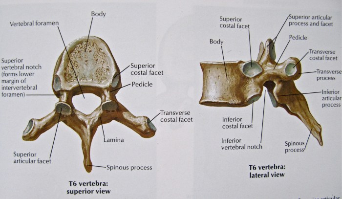
Each spinal bone has a unique shape and structure that allows it to perform specific functions. The bodies of the spinal bones are relatively large and flat, providing support for the weight of the upper body. The spinous processes, which are projections at the back of the spinal bones, provide attachment points for muscles and ligaments that help to stabilize the vertebral column.
The spinal bones also have a series of holes called foramina, which allow for the passage of nerves and blood vessels. The spinal cord, which is a bundle of nerves that carries messages between the brain and the rest of the body, passes through the vertebral canal, which is formed by the foramina of the spinal bones.
Types of Spinal Bones

The spinal column, also known as the backbone, is a complex structure composed of 33 individual bones known as vertebrae. These vertebrae are classified into five distinct types based on their location and specific characteristics: cervical, thoracic, lumbar, sacral, and coccygeal.
Cervical Vertebrae
The cervical vertebrae are located in the neck region and are the most mobile of all the spinal bones. They are characterized by a small, triangular shape with a large opening for the spinal cord. The first cervical vertebra, known as the atlas, is unique in that it articulates with the skull and allows for head movement.
The second cervical vertebra, the axis, has a prominent odontoid process that articulates with the atlas, facilitating rotation of the head.
Thoracic Vertebrae
The thoracic vertebrae are located in the chest region and are larger and more robust than the cervical vertebrae. They have a more rectangular shape and feature facets for articulation with the ribs. The thoracic vertebrae provide support for the rib cage and protect the vital organs within the chest cavity.
Lumbar Vertebrae
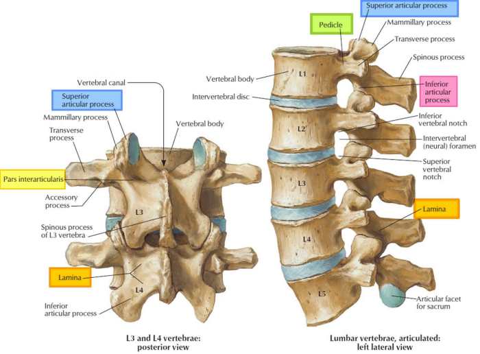
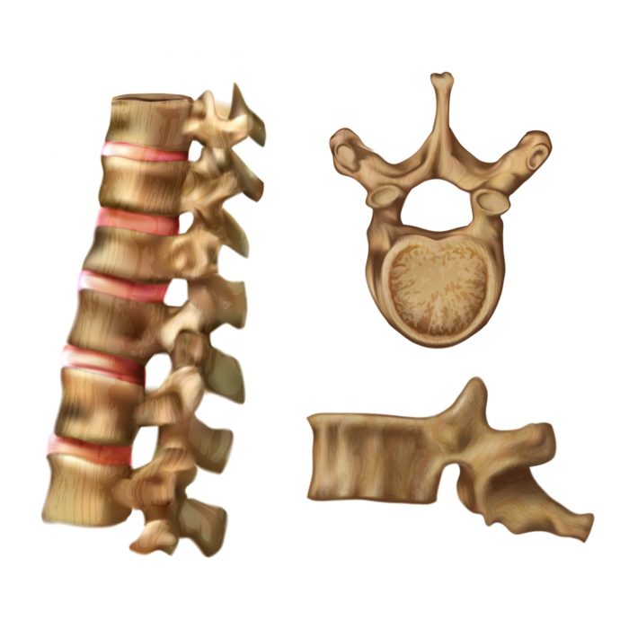
The lumbar vertebrae are located in the lower back region and are the largest and strongest of the spinal bones. They are characterized by a massive, kidney-shaped body and have thick pedicles and laminae. The lumbar vertebrae bear the weight of the upper body and provide stability during movement.
Sacral Vertebrae
The sacral vertebrae are located in the pelvic region and are fused together to form a single, triangular bone known as the sacrum. The sacrum articulates with the hip bones and provides support for the pelvic organs. It also serves as an attachment point for ligaments and muscles that stabilize the pelvis.
Coccygeal Vertebrae
The coccygeal vertebrae are located at the base of the spine and are the smallest and least developed of the spinal bones. They are typically fused together to form a small, triangular bone known as the coccyx. The coccyx provides minimal support and is often referred to as the “tailbone.”
Structure of Spinal Bones: Any Of The 12 Spinal Bones Crossword
The spinal bones, also known as vertebrae, are stacked upon one another to form the spinal column. Each vertebra has a unique structure that contributes to the overall stability and flexibility of the spine.
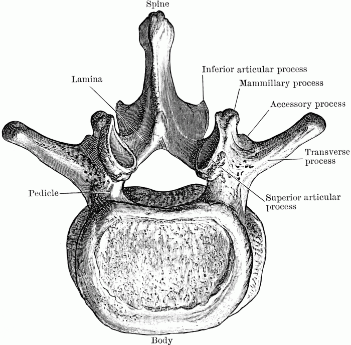
A typical spinal bone consists of several key components:
Vertebral Body
- The vertebral body is the large, cylindrical front portion of the vertebra. It bears the weight of the body and provides structural support.
- The upper and lower surfaces of the vertebral body are covered with cartilage, which allows for smooth movement between the vertebrae.
Pedicles
- The pedicles are two short, thick projections that extend from the back of the vertebral body.
- They connect the vertebral body to the laminae.
Laminae
- The laminae are two flat plates that extend from the pedicles and meet at the back of the vertebra.
- They form the roof of the vertebral foramen, which is the opening through which the spinal cord passes.
Processes, Any of the 12 spinal bones crossword
- The processes are various bony projections that extend from the vertebrae.
- They serve as attachment points for muscles, ligaments, and tendons.
- The most prominent processes are the transverse processes, which extend laterally from the vertebrae, and the spinous process, which projects posteriorly.
Articulations of Spinal Bones

The spinal bones, also known as vertebrae, articulate with each other to form the spinal column. These articulations allow for movement and flexibility of the spine while providing stability and support.
There are three main types of joints involved in the articulation of spinal bones:
Intervertebral Discs
- Intervertebral discs are fibrocartilaginous pads located between adjacent vertebrae.
- They act as shock absorbers, cushioning the spine during movement.
- The intervertebral discs also provide flexibility and allow for a limited range of motion between vertebrae.
Facet Joints
- Facet joints are synovial joints located on the posterior aspect of adjacent vertebrae.
- They allow for gliding and rotational movements of the spine.
- The facet joints are lined with cartilage and surrounded by a joint capsule, providing stability and preventing excessive movement.
Clinical Significance

The spinal bones play a crucial role in maintaining the structural integrity of the body and protecting the delicate nervous system. Understanding their clinical significance is essential for diagnosing and treating various spinal conditions.
Spinal injuries can have a profound impact on the nervous system and overall health. Trauma, degenerative diseases, or congenital abnormalities can damage the spinal cord or nerve roots, leading to neurological deficits such as paralysis, sensory loss, and autonomic dysfunction.
Spinal Injuries and Neurological Effects
Spinal injuries can result in a wide range of neurological impairments depending on the severity and location of the damage. These may include:
- Paralysis:Loss of motor function below the level of injury, resulting in weakness or complete loss of movement.
- Sensory loss:Impaired sensation, including touch, pain, temperature, and proprioception, in areas innervated by the affected nerve roots.
- Autonomic dysfunction:Disruption of involuntary bodily functions, such as blood pressure regulation, bladder and bowel control, and sexual function.
The severity and prognosis of spinal injuries depend on factors such as the type and extent of damage, the level of the injury, and the patient’s overall health.
General Inquiries
What is the primary function of the spinal bones?
The spinal bones serve as the foundational pillars of our skeletal framework, providing support and stability to the body while safeguarding the delicate spinal cord within.
How many types of spinal bones are there?
The spinal column comprises five distinct types of spinal bones: cervical (neck), thoracic (chest), lumbar (lower back), sacral (pelvis), and coccygeal (tailbone).
What are the potential consequences of spinal injuries?
Spinal injuries can have severe implications, ranging from localized pain and discomfort to neurological impairments and even paralysis. Protecting the spinal column is paramount for maintaining optimal health and mobility.